Microbes are microscopically small living organisms. The small size of the cells, which have a very large surface area relative to their mass, results in lively metabolic activity, so leading to rapid cell enlargement and multiplication and a high level of cell respiration. They are capable of surviving unfavorable living conditions (e.g. unfavorable temperatures) by forming spores. If living conditions then become favorable again, they germinate. Thus, for example, if a container has not been carefully cleaned, goods may subsequently become infected with spores from previous cargoes which resume activity in the new environment (secondary infection). This is why refrigerated containers must be in a thoroughly hygienic condition.
Fungal molds form hyphae. i.e. branched fungal filaments, which are visible as an absorbent cotton-like covering (mycelium) and break up the surface of the substrate or make it slimy. The actual layer of mold is formed by the conidiophores, which produce the spores (conidia). The development of fungal molds is divided into four phases, damage to goods occurring in the third and fourth phases:
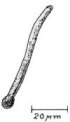
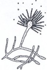
| 1st phase | - | Germination of an unbranched mycelium fiber (Fig. 68) |
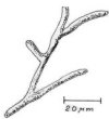
| 2nd phase | - | Formation of the original mycelium, which grows and forms four to five hyphae (Fig. 69) |
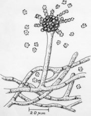
| 3rd phase | - | Developed mycelium with numerous hyphae and a number of fruiting bodies (Fig. 70, Fig. 74) |
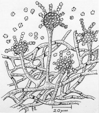
| 4th phase | - | Reproductive stage with very dense mycelium layers and numerous conidiophores with spores (Fig. 71, Fig. 75). |
 |
 |
| Figure 68: Stage I of mycelial growth |
Figure 69: Stage II of mycelial growth |
 |
 |
| Figure 70: Stage III of mycelial growth |
Figure 71: Stage IV of mycelial growth |
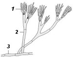 1 - Conidia 2 - Conidiophore 3 - Mycelial filament |
 |
| Figure 72: Penicillin mold (Penicillium italicum) [29] |
Figure 73: Aspergillus flavus [34] |
Figs. 72 and 73 show two important examples of fungal molds, penicillin mold (Penicillium italicum) and Aspergillus flavus; the latter forms aflatoxin which is one of the most potent cytotoxins which has yet been discovered.
 |
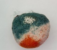 |
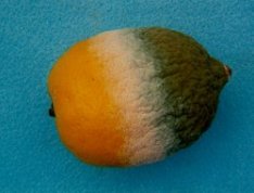
| Figure 74: Lemon, suffering from green mold - stage III (white) and stage IV (green) | Figure 75: Blue mold caused by the fungal mold Penicillium italicum, stage III (white) and stage IV (blue), on an orange. Spores can be seen at the top. |
Fig. 76 shows fresh ginger, which began to go moldy (white) and to sprout (green) in a refrigerated container because the temperature and humidity were too high.
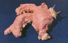 |
Figure 76: Fresh ginger, which began to go moldy (white) and to sprout (green) in a refrigerated container because the temperature and humidity were too high |